
From Soft Scaffolding to Sturdy Support: Unveiling the Wonders of Bone Ossification
A Deep Dive into Intramembranous and Endochondral Ossification
A Deep Dive into Intramembranous and Endochondral Ossification
Have you ever wondered how your bones, the sturdy framework that supports your body, come into existence? This fascinating process, called bone ossification, starts even before you're born and continues well into your teenage years. It's a captivating story of cellular transformation, orchestrated by specialized cells, that sculpts your skeleton from a soft, jelly-like substance.
This article delves into the two main types of bone ossification: intramembranous and endochondral ossification. We'll explore how these processes work, their clinical relevance, and the amazing cellular teamwork that brings them to life.
The Direct Approach: Intramembranous Ossification
Imagine building a house directly from bricks – that's the essence of intramembranous ossification. Here, bone formation happens within a specialized tissue called mesenchymal connective tissue, which acts as the building block for various structures in the body.
This process begins in the womb and extends into adolescence. It's responsible for forming flat bones like the skull and clavicle (collarbone). Interestingly, the skull isn't fully ossified at birth. This allows the head to compress slightly during childbirth, making passage through the birth canal smoother for both mother and baby.
Let's zoom in and see the cellular magic at play:
- Stem Cell Symphony: Mesenchymal stem cells, the masters of cellular transformation, kick things off. They differentiate (transform) into various specialized cells, including blood vessel builders and the stars of the show – osteoblasts.
- Osteoid Orchestra: Osteoblasts, the bone-building maestros, take center stage. They secrete a protein-rich material called osteoid, the unmineralized foundation of future bone.
- Mineral Mania: As osteoid accumulates, calcium phosphate crystals bind to it, hardening the matrix and transforming it into true bone. Some osteoblasts become trapped within this mineralized matrix, morphing into osteocytes – the bone's maintenance crew.
- Double Duty Duo: Meanwhile, on the bone's surface, the periosteum, a tough membrane, plays a dual role. Its inner layer harbors osteogenic cells, which mature into osteoblasts, continuously adding new bone layers. The outer layer anchors muscles and tendons, providing stability.
The Cartilage Canvas: Endochondral Ossification
This process takes a more roundabout route, using a temporary cartilage model as a blueprint for bone formation. It's responsible for crafting your long bones like the femur (thigh bone) and humerus (upper arm bone).
The story begins even earlier, around 6-8 weeks after conception:
- Hyaline Haven: Mesenchymal cells morph into chondroblasts, which churn out hyaline cartilage, a flexible yet strong tissue. This cartilage model serves as the initial bone template.
- Cartilage Calcification: As development progresses, chondrocytes (cartilage cells) in the center of the model enlarge and eventually die. This triggers the calcification of the surrounding matrix, making it hard.
- Blood Vessel Bonanza: The perichondrium, a membrane surrounding the cartilage, transforms into the periosteum. This crucial step brings in blood vessels carrying osteoblasts, the bone-building champions.
- Bony Collar: Osteoblasts get to work, depositing bone around the shaft of the developing bone, forming a collar. This cuts off nutrient supply to the central cartilage, leading to its degeneration.
- Primary Ossification Powerhouse: Blood vessels invade the now-hollow spaces left behind by the degenerating cartilage, carrying osteoblasts with them. These osteoblasts establish the primary ossification center, the core of future bone growth.
- Growth Plate Grandeur: Meanwhile, the cartilage model continues to grow at its ends. This growth zone is called the epiphyseal growth plate. Here, chondrocytes continuously multiply, pushing the bone ends further apart, allowing the bone to lengthen.
- Secondary Surprise: After birth, secondary ossification centers appear at the ends of the long bones, following a similar process to the primary center.
- Epiphyseal Farewell: As you reach adulthood, the growth plate matures, and cell division ceases. Eventually, the growth plate itself ossifies, fusing the epiphysis (bone end) to the diaphysis (bone shaft), marking the end of bone growth in length. The only remaining cartilage is found in the smooth articular surfaces of your joints, enabling pain-free movement.
The Body's Blueprint: Clinical Relevance of Ossification
Understanding bone ossification is crucial in various medical contexts:
- Bone Age Assessment: By analyzing X-rays of bones with growth plates, doctors can estimate a child's skeletal maturity. This helps assess growth patterns and identify potential developmental delays or hormonal imbalances.
- Growth Plate Injuries: The growth plate, while crucial for bone lengthening, is also a vulnerable spot. Fractures in this area require prompt medical attention to prevent complications like limbs of unequal length. A classification system like the Salter-Harris system helps categorize these fractures for appropriate treatment.
- Bone Disorders: Understanding ossification is vital in diagnosing and managing bone disorders like achondroplasia, a genetic condition affecting cartilage development, and osteogenesis imperfecta, characterized by bone fragility due to collagen defects.
- Bone Regeneration: Research on ossification paves the way for advancements in bone regeneration techniques. Scientists are exploring ways to stimulate osteoblast activity and promote bone growth for treating fractures, bone defects, and age-related bone loss.
Beyond the Basics: A Look Ahead
The fascinating world of bone ossification is constantly evolving. Researchers are delving deeper into the intricate cellular communication pathways that orchestrate bone formation. Additionally, the potential of stem cells to differentiate into bone-building cells holds promise for future bone repair and regeneration therapies.
As we explore further, the remarkable journey of bone formation, from its humble beginnings in the womb to the sturdy skeleton that supports us throughout life, continues to inspire awe. This intricate biological marvel is a testament to the body's incredible ability to build and maintain itself.
About the Creator
suren arju
Hi there! I'm Suren, your startup guide. Entrepreneur, writer, dreamer - I share insights, tips & stories to fuel your startup journey. Ready to explore, learn & win together? Join me & let's redefine how we launch, learn & leap!
Enjoyed the story? Support the Creator.
Subscribe for free to receive all their stories in your feed. You could also pledge your support or give them a one-off tip, letting them know you appreciate their work.
Keep reading
More stories from suren arju and writers in Longevity and other communities.
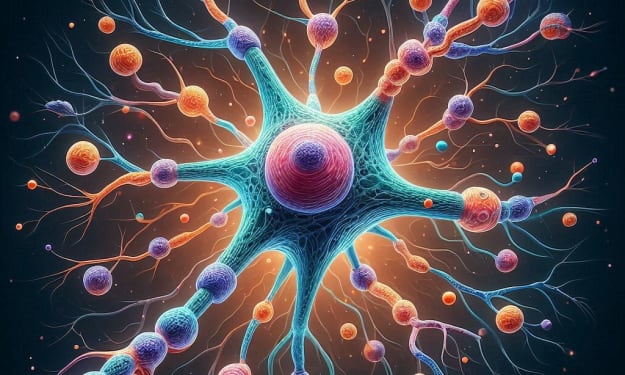
The Symphony Within: Unveiling the Secrets of Nerve Communication
The Hidden Symphony Within: Unveiling the Marvels of Nerve Cell Communication Imagine a bustling metropolis, a symphony of activity orchestrated by a complex network. This isn't a city of steel and glass, but the intricate world within your body – your nervous system. Billions of nerve cells, called neurons, act as the city's communication network, sending and receiving electrical signals that govern everything from muscle movement to the subtlest emotional nuance. But how do these microscopic marvels achieve such remarkable feats? Let's delve into the fascinating ultrastructure of nerves, exploring their building blocks and the intricate dance that allows us to think, feel, and move.
By suren arju5 days ago in Longevity
Scared of Surgery Anesthesia? You Can Cure Yourself of that Awful Fear
A friend recently published a story about their fear of anesthesia and surgical procedures. They said they felt like a coward. I am here to say they are not cowardly—they are human and normal. We are all anxious and intimidated when we face serious medical issues.
By Maryan Pellandabout a month ago in Longevity
The Great Uncoupling of 2229
“I realize there are still aspects of humanity that my kind cannot understand or appreciate….” The soft, airy voice resonating from the orb that trailed Teyhx through the hallway of her dorm pleaded with her to see reason. Though, ultimately, the decision was not hers anyways.
By Adam Clost2 days ago in Futurism


Comments (1)
Love Biology! Great article.