
What are The 2 Main Stages of Thyroid Eye Disease
What is thyroid eye disease?
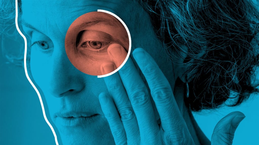
Thyroid eye disease (TED), also known as thyroid-associated orbitopathy and Graves ophthalmopathy is a common orbital disorder. It is one of the most common causes of unilateral and bilateral proptosis in adults. It is an autoimmune disease associated with the activation of antibodies against the thyroid receptors. As a result, excessive thyroid hormone is released leading to the manifestation of hyperthyroidism in different organs of the body. The severity of the disease depends on the stages of thyroid eye disease.
Pathophysiology:
How thyroid eye disease develops?
The most common cause of hyperthyroidism is Graves disease. In this disease, the IgG antibodies bind to the thyroid stimulating hormone (TSH) receptors in the thyroid gland. As a result, thyroid hormone is released.
Thyroid eye disease symptoms:
What are the symptoms of thyroid eye disease?
The most common age group of hyperthyroid is the fourth and fifth decade. The common presentation includes weight loss, increased bowel frequency, sweating, heat intolerance, nervousness, irritability, palpitation, weakness, and fatigue.
Thyroid eye disease signs:
What are the signs of thyroid eye disease?
Signs of hyperthyroidism include thyroid gland enlargement, tremors, palmar erythema, warm and sweaty skin, thyroid acropachy (clubbing of fingers), pretibial myxedema, and indurated skin of shins. Cardiac manifestation includes sinus tachycardia and other arrhythmias.
The treatment of hyperthyroidism includes carbimazole, propylthiouracil, propranolol, thyroid ablation with radioactive iodine, and partial thyroidectomy.
Risk factors for thyroid ophthalmopathy:
Which is the affected population in thyroid eye disease?
• Once a patient develops graves disease, the major clinical risk factor for developing thyroid eye disease is smoking. The greater the number of cigarettes smoked per day, the greater the risk of developing thyroid eye disease. However, quitting smoking can reduce the risk.
• Women are ten times more affected with TED than men. It reflects the fact that women are more affected with Graves disease too. It is more common in middle age population. As TED is an autoimmune disease, any family member affected with the disease makes the family line prone to this disease. The prevalence of the disease is not exactly known but is estimated to be 16 per 100,000 women in the general population and 2.9 per 100,000 men in the general population.
• Radioactive iodine used in the treatment of hyperthyroidism can cause thyroid eye disease.
• Thyroid eye disease can less commonly occur in euthyroid and hypothyroid patients.
I have written an article about some rare eye diseases and read that too.
Clinical course:
How thyroid eye disease proceeds?
Two main stages of thyroid eye disease.
• Inflammatory stage:
The inflammatory stage is also labeled as a congestive stage. It consists of red and painful eyes. The stage lasts up to 1 to 3 years. Only 10% of patients develop serious eye conditions.
• Fibrotic stage:
The fibrotic stage is also known as the quiescent stage. In this stage, the eye is white and has a painless motility defect.
Complications:
What are the complications of thyroid eye disease?
1. Lid retraction:
Lid retraction of the upper and lower lid is present in 50% of the patients and is the most common sign of thyroid eye disease. It is due to the overstimulation of the sympathetic nervous system induced by higher levels of thyroid hormones. Fibrotic contracture of the levator palpebrae and inferior rectus muscle is another postulated mechanism.
Signs include:
• Stellwag sign- Incomplete and frequent blinking
• Dalrymple sign- is lid retraction in primary gaze.
• Kocher sign- it describes staring and the frightening appearance of the eyes
• Enroth sign- lower eyelid edema
• Griffith sign- Lid lag on up gaze
• Von Graefe sign- it signifies retarded descent of upper lid on downward gaze.
• Proptosis:
Proptosis is the outward bulging of the eyes. It is axial, unilateral or bilateral, symmetrical or asymmetrical, and frequently permanent. Severe proptosis compromises the eyelids' normal function, e.g., infrequent blink rate and tear dysfunction leading to exposure keratopathy, corneal ulceration, and infection.
• Restrictive myopathy:
Restrictive myopathy may be permanent. it develops in 30% to 50% of patients with thyroid eye disease. The most commonly affected muscle is the Inferior rectus followed by the medial, superior, lateral rectus, and oblique. The affected muscles result in ocular misalignment, diplopia, and Inability to look up.
• Optic neuropathy:
It is a severe complication of TED. It is caused by compression of the optic nerve or its blood supply at the orbital apex. It results in congestion and enlargement of the recti and orbital tissues. The signs of optic nerve damage include a decrease in vision, color vision, contrast sensitivity, and relative afferent papillary defect. The characteristic visual fields commonly show central, cecocentral, paracentral, and nerve fiber layer bundle defects.
Investigations:
How to test for thyroid eye disease?
• Blood tests for thyroid eye disease include thyroid function tests. Thyroid function is commonly tested initially with TSH, if it is low or normal and still thyroid disease is suspected, additional investigations can be carried out.
• Visual field testing for suspicion of optic nerve compression.
• MRI, CT, and ultrasound are performed for the identification of the typical appearance of extraocular muscles. The muscle belly enlargement with tendon sparing is a typical sign of TED.
• The visual evoked potential is sometimes used in optic neuropathy.
Clinical Activity Scoring:
The stages of thyroid eye disease can be calculated with the European Group on Graves’ disease orbitopathy (EUGOGO). The severity scoring helps to keep a low threshold for the use of immunosuppressants. One point is given to each of the following
• Spontaneous orbital pain
• Gaze evoked orbital pain
• Eyelid swelling (due to inflammatory phase)
• Eyelid erythema
• Conjunctival redness (due to the inflammatory phase)
• Chemosis
• Inflammation of caruncle or plica.
1. Mild disease: Thyroid eye disease is labeled as a mild disease when there is only a minor impact on daily life.
2. Moderate disease: In moderate eye disease, there is soft tissue involvement, a lid retraction of 2mm or more, and diplopia and proptosis of 3mm or more.
3. Severe disease: The disease is labeled as severe when there is corneal involvement or optic neuropathy.
Management
How do they treat the thyroid eye?
Depending on the stages of thyroid eye disease, the treatment begins with the cessation of smoking and controlling thyroid dysfunction. If the patient has previously used radioiodine for the treatment of hyperthyroidism, a short course of oral steroids should be instituted.
Mild disease:
• In mild diseases, lubricants should be used for corneal exposure and dryness.
• The use of topical steroids and anti-inflammatory drugs is advocated by some authorities
• Periorbital edema can be reduced by keeping the pillows elevated during sleep time.
• Taping of the eyelids during sleep can reduce damage brought by exposure to keratopathy.
Moderate disease:
Systemic steroid therapy is the mainstay of treatment for moderate to severe thyroid eye disease.
• Oral prednisolone can be given initially with a dose of 60-80 mg/day and tapered gradually.
• Lower-intensity doses of intravenous steroids can be given in absence of acute sight-threatening disease. The dose is 0.5 g once weekly for 6 weeks followed by 0.25 g for another 6 weeks. The symptoms start to improve within 24 hours with a reduction in discomfort, chemosis, and periorbital edema with a maximum response within 2-8 weeks.
• Orbital steroids are seldomly used with a minimum side effect but are considered less effective than systemic treatment.
• Low-dose fractioned radiotherapy can be used along with steroids or when steroids are ineffective or contraindicated. The effect is much delayed as compared to steroids. A positive response is usually noticed within 6 weeks with maximum improvement in 4 months. The adverse effects include cataracts, radiation retinopathy, optic neuropathy, and an increased risk of local cancer. The threshold should be high in young patients and diabetics because of the increased of retinopathy.
• Combined therapy with irradiation, azathioprine, and low-dose prednisolone may be more effective than steroids or radiation alone.
Severe disease:
Optic neuropathy and exposure keratopathy is the severe stage of thyroid eye disease. It needs aggressive therapy.
The therapy is started as intravenous prednisolone with a dose of 0.5-1.0 g on three successive days with conversion to the oral treatment of 0.5- 1.0 g on alternative days, 3-6 times, keeping the daily dose lower than 8 g to avoid the complication of liver compromise.
Post-inflammatory complications/ surgery to treat TED:
Post-inflammatory complications develop after the inflammatory phase has subsided. These includes
1. Proptosis: After the active inflammatory phase has been remitted, the patient may be rendered with functional or cosmetic proptosis. Surgical decompression is the choice of surgery, to reduce the proptosis. In this procedure, the bony walls of the orbit are removed so that volume is increased in the orbit. This may be one-wall, two-wall, or three-wall procedures. The complications include hypoglobus, diplopia, and damage to the infraorbital nerve.
2. Restrictive myopathy: Surgery is usually performed in patients with diplopia in the primary or reading gaze, provided that the inflammatory phase has subsided and the angle of deviation is stable for 6-12 months. The aim of the surgery is to gain single vision in primary and reading gaze. The recession of the inferior or medial rectus is the commonly performed procedure.
3. Lid retraction: Mild ptosis may improve spontaneously or with the control of hyperthyroidism. However, some cases may require surgical intervention. It includes mullerectomy for mild cases while recession or disinsertion of levator aponeurosis may be needed in severe cases. The recession of lower lid retractors, with or without hard palate graft can be performed with lower lid retraction of more than 2mm.
FDA approves first treatment for thyroid eye disease
For Details Click Here
About the Creator
sadia ayaz
My name is Dr. Sadia Ayaz and I am an ophthalmologist and eye surgeon. My medical degree is from Pakistan, I have done FCPS-1 / IMM from the College of Physicians and Surgeons Pakistan.
Enjoyed the story? Support the Creator.
Subscribe for free to receive all their stories in your feed. You could also pledge your support or give them a one-off tip, letting them know you appreciate their work.
Keep reading
More stories from sadia ayaz and writers in Longevity and other communities.
Macular Hole Stages: Protect Your Vision with Early Intervention
What is Macular Hole? The macular hole is a defect or lesion extending from ILM (internal limiting membrane) deep down to the RPE (retinal pigment epithelium). As the macula is responsible for keeping the central vision intact. Any pathology at the region of macular will lead to central vision loss. Depending upon the macular hole stages, the severity of vision impairment can be graded. It is a condition in which fellow eye can be involved with a risk of other eye involvement at 5 years in around 10% cases The article will discuss the causes, signs and symptoms, macular hole stages and available treatment options, prevention, outlook and prognosis.
By sadia ayazabout a year ago in Longevity
Takeaways from 2 Years of Duolingo Spanish
Two years ago on this date, I downloaded the Duolingo app for approximately the 15th time. When I’d been inspired to try brushing up on my Spanish in the past, I’d start out hot and heavy only to sputter out after a few weeks of enthusiasm and dedication.
By Veronica Wren26 days ago in Longevity


Comments
There are no comments for this story
Be the first to respond and start the conversation.